
As most of us are aware, an X-ray of the breast is commonly known as a mammogram. But did you know that there are two distinct types of mammograms? There are several different types, but they all fall under two primary categories, which are screening and diagnostic. That is why we think it is important to compare screening vs diagnostic mammogram exams. Here are some of the differences between screening and diagnostic mammogram exams.
Screening vs Diagnostic Mammogram
First of all, screening mammograms are for women who are at average risk of developing breast cancer and do not present any symptoms. The American Cancer Society recommends that all women begin to have such screenings annually beginning at age 40, or each year after age 30 if they have a genetic syndrome that puts them at a higher risk for breast cancer.[1] If a first-degree relative (mother or sister) suffered from the disease, you should start getting screening mammograms at the age that is ten years younger than the diagnosed age of your relative.
Your first screening will be the baseline to compare all future screenings with. This means any differences in tissue should appear obvious. Because of this, you must be sure to share any previous mammograms done in a different facility with your current provider. It is always best to allow the radiologist to compare past and present mammograms. You can usually pick them up yourself and take them to your appointment or sign a release with both offices so they can exchange otherwise HIPAA-protected information.
If during a screening mammogram there is something spotted that is indeterminable or blatantly suspicious, you will get a referral to get a diagnostic exam.
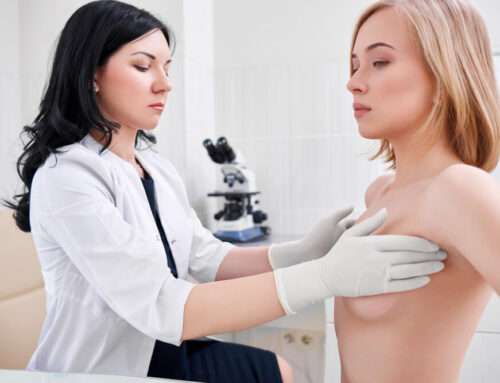
This mammogram diagnostic, as the name implies, seeks to diagnose abnormal findings seen on the initial screening. These may include some signs of breast cancer that would alert an astute doctor and push him or her to further investigate. Some common symptoms include lumps, changes in the shape and size of breasts, and thickening of breast skin and tissue. In addition, you may be experiencing nipple discharge and breast pain. All findings will be thoroughly examined to determine if they are indicative of a breast cancer diagnosis.
If you had prior treatment for breast cancer or had a previous diagnostic exam, you may also receive a mammogram diagnostic procedure during your follow-up visits.
Each difference between screening and diagnostic mammogram exams is extreme, but they do have a lot in common. They both utilize either three-dimensional technology called tomosynthesis or rely on the typical two-dimensional mammography in a digital format.
How Screening Mammograms Work
The Centers for Disease Control acknowledges a screening mammogram as the easiest and most effective way to detect breast cancer in its earliest stages.[2] Mammograms can detect lumps that are not detectable by the woman herself. Finding out early gives you the best prognosis because you have the most treatment options available.
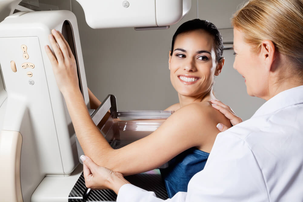
A screening mammogram simply checks the breast for any signs of cancer. Having a comprehensive screening exam ensures that your physician will immediately find, diagnose, and, if needed, treat any suspicious changes in breast tissue. It is a precautionary measure taken annually by women that are not experiencing any symptoms.
The process usually takes no longer than ten to fifteen minutes and may be a bit uncomfortable, though not in any way invasive. Your physician does not have to be present during the exam, though they will interpret the results afterward.
How Diagnostic Mammograms Work
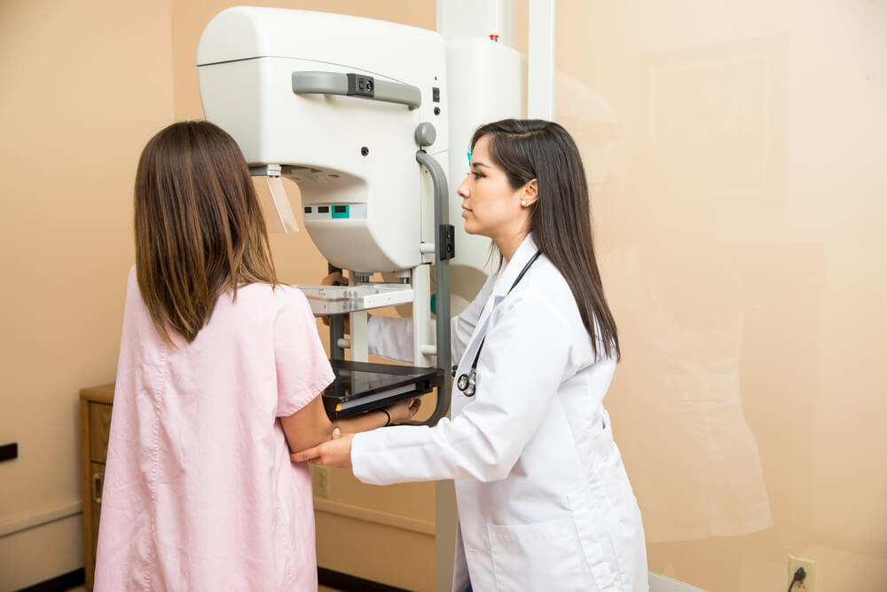
Diagnostic mammograms provide a much more comprehensive look at the breast than a regular screening mammogram would. This is due to the special techniques they employ to get a more detailed picture. They also are helpful for screening women with breast implants who are difficult to examine.
If you’re referred for a diagnostic exam, you need to know that it is a much longer process than a screening exam. This is because there will be multiple X-rays taken from several viewpoints of the breasts in order to get a holistic picture of an anomaly. The radiologist is also likely to zoom in on the abnormal tissue to get a clear image for the doctor to examine. Short of a biopsy, this is his or her best tool for determining a diagnosis.
In addition to masses, diagnostic mammograms have been crucial in the finding of spot ductal carcinoma, or DCIS, along the breast duct. The primary sign of DCIS is microcalcifications, which are abnormal cells that are similar to small grains of sand, and they are extremely difficult to detect. When these cells group closely together in a row, they can become a serious, invasive cancer.
Your physician will usually be present during a diagnostic mammogram. X-rays are usually reviewed immediately since time is imperative when fighting breast cancer. Usually, you will wait in the office during this review, so you will be available in case your doctor requires extra images.
Other technologies, such as breast MRIs and the ABUS whole-breast ultrasound, are also procedures used to expand on the mammogram’s findings.
Sometimes, a magnification view or a spot view, which are special images, will be necessary to further examine a specific, troubling area. A breast ultrasound can assist the doctor with interpreting what they spotted on the mammogram.
A diagnostic mammogram may reveal that tissue that seemed concerning on your screening exam is perfectly fine after all. If this occurs, you simply return to getting a screening exam each year.
Sometimes, a suspicious area is cancer-free, but your doctor might have you come back in six months to keep an eye on it. This is because they want to watch it carefully.
If a suspected area appears to be breast cancer, your doctor may need to schedule a biopsy to confirm their suspicion. However, just because they referred you for a biopsy does not mean that you have cancer. It is just a precautionary measure.
The Reliability of Mammograms
The density of your breasts’ tissue and the size of any tumors are pertinent to the success of a mammogram in finding breast cancer. It is also crucial that you have a skilled radiologist performing the mammogram and a proficient doctor reading it over.
There are other factors to consider, too. Breast tumors in women under age fifty are harder to find. This is likely due to the fact that dense breast tissue simply looks white on the X-ray, as do tumors, and younger women tend to have denser breasts.
This is why three-dimensional mammography is the best approach for the highest percentage of accuracy. As a matter of fact, this technology has a reputation for detecting breast cancer 28% more accurately than analog X-rays.
Health Insurance Coverage
The Affordable Care Act has made it a requirement that all new health insurance plans fully cover screening mammograms. This means that, since 2010, there have been free screening mammograms. Plus, if you are on Medicare and over forty, your screening exams are fully covered.

Unfortunately, diagnostic mammograms are not completely covered like free screening mammograms. They fall under the terms unique to private and employer-based insurance plans. You would have to look up how your plan covers screening vs diagnostic mammogram exams with your human resources department.
In conclusion, screening mammograms are a preemptive tool, and diagnostic mammograms are a closer look at anything that may concern your physician. Both are crucial and effective tools that save many women’s lives every year. So, if you have not had your annual screening, it is imperative that you schedule yours today with the radiologists at Breast Care Center Miami. Come into our office or call us today to schedule your appointment.
Do you have questions? Comment below, and we will answer you as best as we can.
References
- https://www.cancer.org/healthy/find-cancer-early/cancer-screening-guidelines/american-cancer-society-guidelines-for-the-early-detection-of-cancer.html
- https://www.cdc.gov/cancer/breast/basic_info/mammograms.htm#:~:text=A%20mammogram%20is%20an%20X,before%20it%20can%20be%20felt.