
Blog
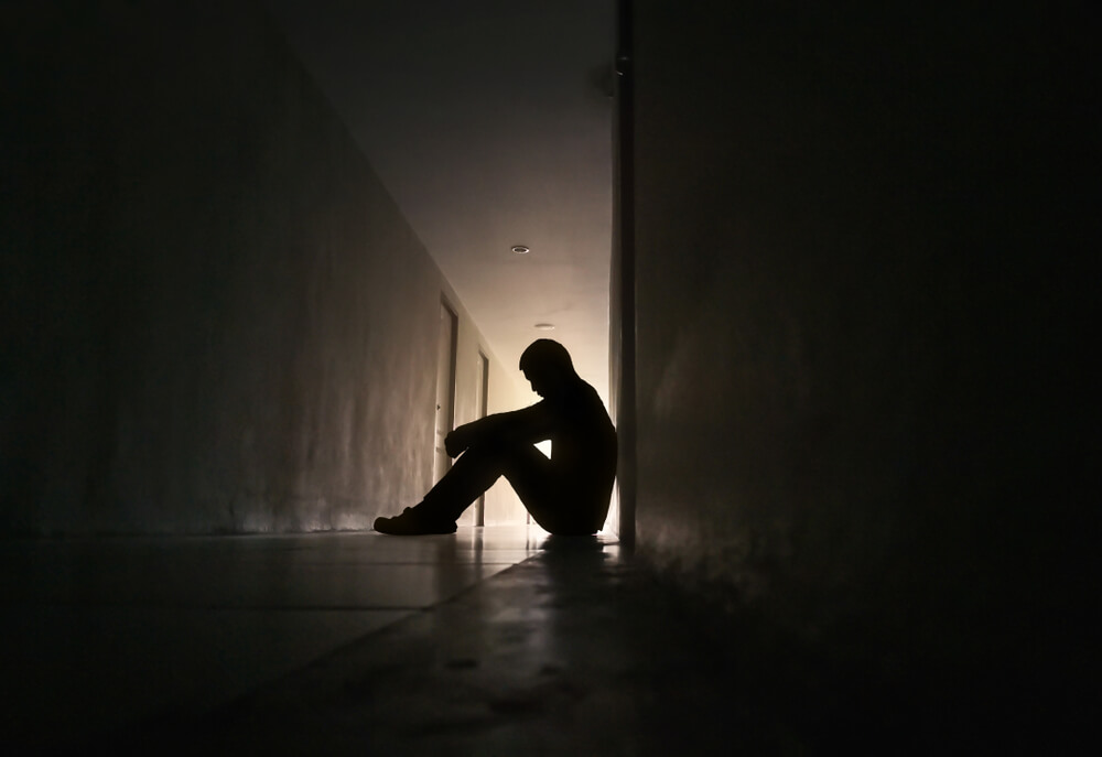
What Does Anxiety Feel Like: Physical Symptoms Of Anxiety
Much of modern life moves at a rapid, unrelenting pace.
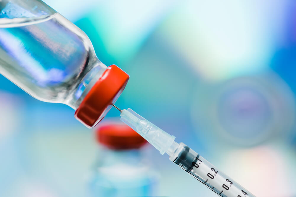
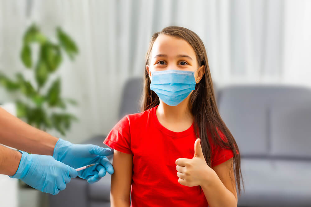
MMR Vaccine: Protection from Measles, Mumps, and Rubella
The MMR vaccine, that stands for Measles, Mumps, Rubella vaccine,
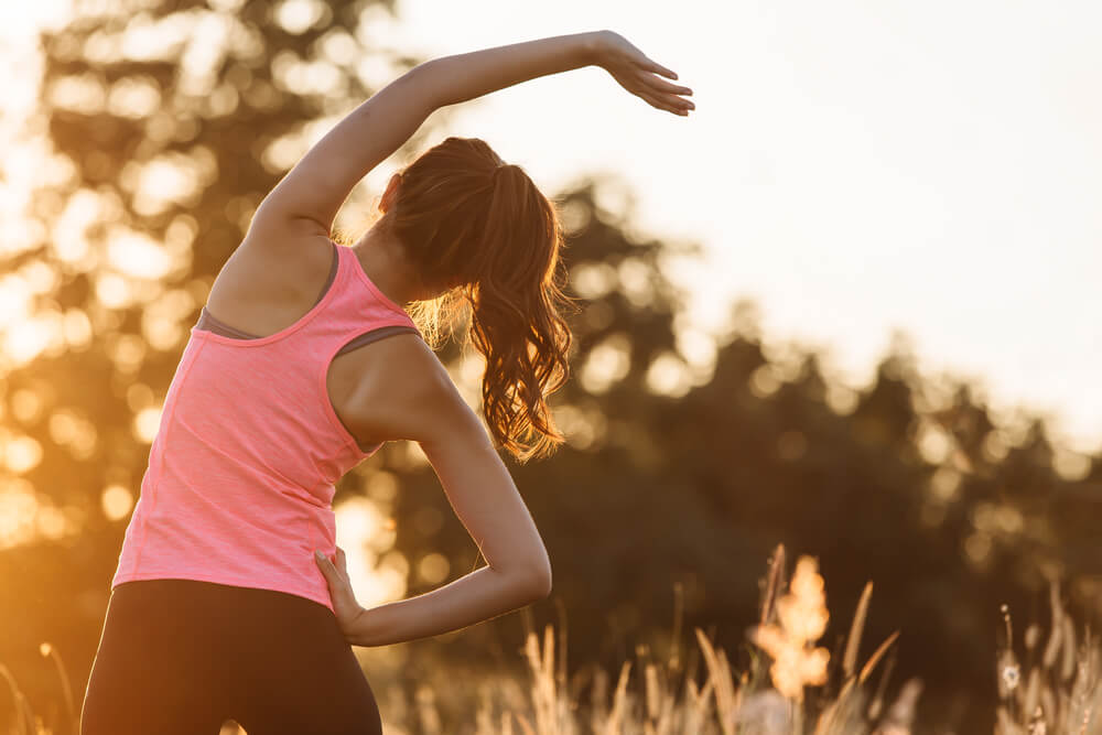
Health & Wellness Tips: Improving Your General Health
In the hustle and bustle of today's fast-paced world, prioritizing

Sedentary Lifestyle: How To Improve Your Lifestyle
A sedentary lifestyle is a kind of living characterized by

Top Essential Questions to Ask Your Doctor
Effective communication is one of the most pivotal aspects of
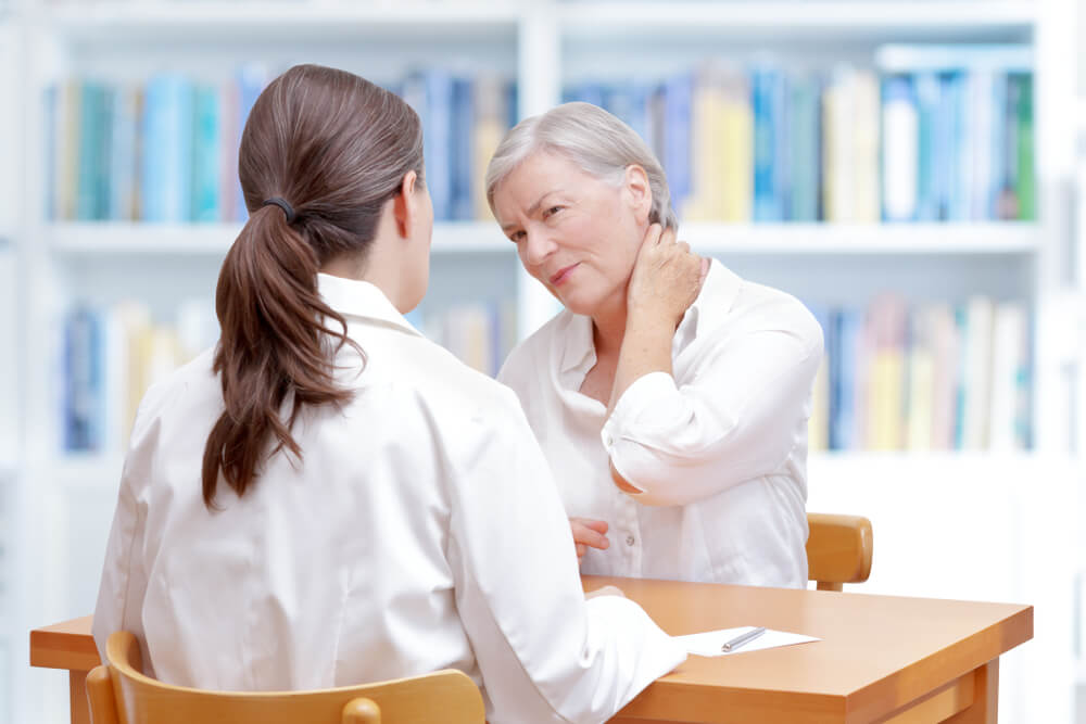
Effective Methods for Living with Chronic Pain
Chronic pain, as its name implies, is a constant, recurring
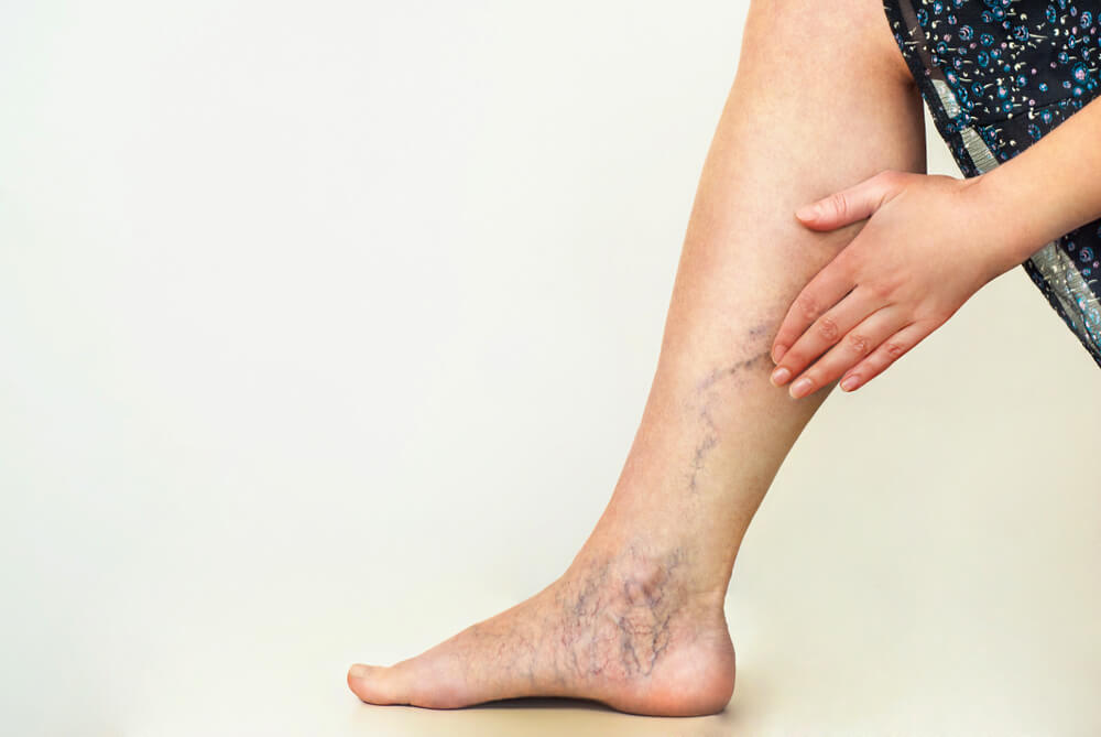
Practical Tips on How to Avoid Varicose Veins – Preventive Guide
Varicose veins, the gnarled, enlarged veins that often appear in
Comprehensive Guide to Varicella Immunization
Varicella, commonly known as chickenpox, is an infectious disease that
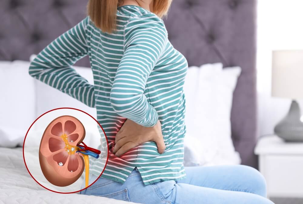
Tips For Passing a Kidney Stone & How To Prevent Them
Experiencing a kidney stone can be a distressing and often