
A prostate ultrasound helps detect abnormalities, diagnose symptoms such as elevated blood test results, and more. But, how does a prostate exam work, and what can you expect along the way? Luckily for you, this comprehensive article will give you all the information you need to prepare for a prostate ultrasound. We will discuss what a prostate ultrasound is, some of the common uses of the procedure, how to prepare, and so on. If this is your first prostate ultrasound, the information in this article will be more than useful.
Of course, besides educating yourself with the facts, it’s also important to seek out professional guidance. If you are located in the area or looking for reliable professionals, we recommend ultrasound in Hialeah, FL.
That said, here’s everything you need to know about a prostate ultrasound. Let us begin.
What is a Prostate Ultrasound?
A prostate ultrasound or a rectal ultrasound is a simple, non-invasive procedure that is performed to diagnose symptoms such as elevated blood test results and urinating difficulties. Moreover, a rectal ultrasound helps investigate a suspicious nodule found during routine rectal exams. The doctor will also check the prostate size for any abnormalities. Overall, a rectal ultrasound is entirely safe, and it does not involve ionizing radiation.
You don’t need to do special preparations for a rectal ultrasound. However, make sure you remove any jewelry and wear comfortable and loose clothing. Doctors may ask you to wear a gown and lie on one side with the knees toward the chest. The doctor will insert a small plastic cylinder or an ultrasound transducer into the rectum to obtain the highest-quality images.
Although a prostate ultrasound is safe and non-invasive, it’s imperative to find a fantastic healthcare provider. If you are looking for the safest option with tons of positive patient testimonials, check out Carreras Medical Center.
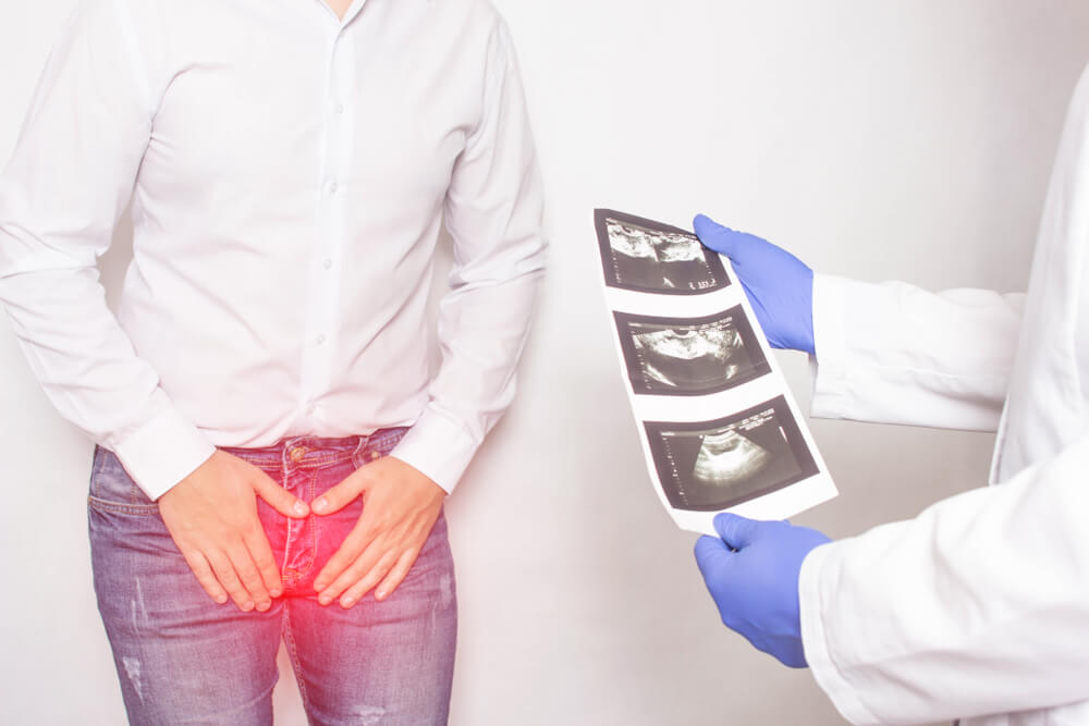
In brief, ultrasound imaging helps professionals diagnose and treat certain medical conditions. A prostate or rectal ultrasound provides quality images of the patient’s prostate gland and surrounding tissue.
Common Uses of the Prostate Ultrasound Procedure

Before you understand the answer to “how does a prostate exam work?”, let’s see the common uses for this procedure. Your doctor may recommend performing a rectal ultrasound to:
- Detect abnormalities within the prostate, such as disordered prostate size.
- Checking whether the prostate size is enlarged (this condition is called BPH or benign prostatic hyperplasia).
- Detect growth within the prostate.
- Help determine the cause of infertility.
Some symptoms that can be diagnosed with the help of a rectal ultrasound include:
- Difficulty urinating.
- Elevated blood test result.
- A suspicious nodule that needs further investigation.
Since a rectal ultrasound provides high-quality, real-time images, doctors can use it to guide procedures like needle biopsies. During needle biopsies, the professional uses a needle to sample cells or tissue in the prostate gland.
How Does a Prostate Exam Work?
Think of ultrasound imaging as the sonar fishermen, ships, and bats use. For instance, when a sound wave hits an object, it echoes or bounces back. By monitoring the echo waves, it’s possible to check the object’s size, consistency, shape, and distance. This principle refers to whether the object is fluid-filled or solid.
Ultrasounds are excellent for identifying changes in the appearance of arteries, tissues, organs, and abnormalities such as tumors.
As mentioned, the doctor will use a transducer carefully inserted into the patient’s rectum. This transducer sends short pulses of high-frequency and inaudible sound waves into the patient’s body. When the sound waves bounce off the interior organs, tissues, and fluids, the sensitive receiver in the transducer records changes in direction and pitch. The computer quickly measures the results of the waves and displays a real-time image on the monitor.
Ultrasound procedures such as rectal ultrasounds perform the same way.
Doctors perform the ultrasound exam transrectally because the prostate gland is in front of the rectum. This way, the imaging probe is close to the gland, and the results can be more accurate.
As mentioned, the healthcare provider will instruct the patient to lie on one side with the knees bent. Next, the expert will cover the lubricated transducer in a protective cover and insert it through the rectum. Finally, in order to gain the best view of the prostate gland, the expert will take images from various angles.
If your healthcare professional finds a suspicious lesion during the rectal examination, you might require an ultrasound-guided biopsy. During this procedure, the professional will insert a needle into the prostate gland while at the same time monitoring the location of the needle. The expert may also want to extract a tissue sample for further microscopic examination.
Moreover, to assess if a patient is at risk for cancer, the doctors may use a PSA or prostate-specific antigen test. In this case, the doctor will perform a biopsy with the help of an ultrasound probe. Usually, a rectal ultrasound will last up to twenty minutes.
What Can I Expect During and Post-Procedure?
Overall, a prostate ultrasound is a simple procedure. Of course, due to its nature, you may experience minimal discomfort. If the patient doesn’t require a biopsy, this type of ultrasound is similar to a regular rectal exam. On the flip side, if the patient needs a biopsy, there may be added discomfort. However, the discomfort is minimal.
In rare cases, there may be small amounts of blood in the urine or sperm post-procedure. When you are done with the ultrasound exam, you can perform most of your normal daily activities. Of course, make sure to take some time off to recover fully.
What are the Pros and Cons of this Procedure?

Just like any procedure, the ultrasound of the prostate can have some benefits and potential drawbacks. Here are some of the most common benefits linked to this procedure:
- It uses zero ionizing radiation.
- It’s widely available, relatively inexpensive, and easy to use.
- It will give you a clear picture of the soft tissues that don’t appear on X-ray images.
- You may repeat an ultrasound as often as needed without causing health problems.
- You get real-time imaging.
If you choose a reputable healthcare provider, your chances of experiencing any adverse effects are zero. However, some unreliable clinics can cause more harm to your health, so choose wisely.
Does this Procedure Have Any Limitations?
Patients who have had the tail end of the rectum removed before surgery are unsuitable candidates for this procedure. The reason for this is that this procedure involves inserting a probe into the rectum. Alternatively, radiologists may try to check the prostate gland by using a standard ultrasound imaging probe on the patient’s perineal skin. In some cases, experts can examine the glands this way, but the results aren’t as detailed as those done with a transrectal probe.
Book an Appointment Today
You deserve to be in excellent shape. If you are concerned about your health, fret not, we’re here. Our team of experts will help get your life back on track. Book an appointment today and get started with your journey.
