![[amniocentesis]](http://cdn1.medicalnewstoday.com/content/images/articles/308/308404/amniocentesis.jpg)
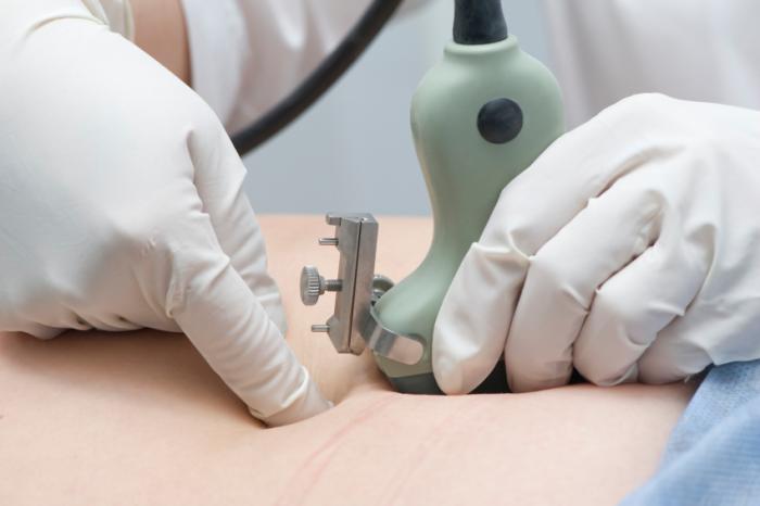
Amniocentesis can detect abnormalities, but is there more to the story?
Normally, there are 23 pairs of chromosomes in each cell of the human embryo: 22 pairs of chromosomes and one pair of sex chromosomes.
Multiple copies of chromosomes in cells can be a sign of developmental disorders. Children who have three copies of chromosome 21 will develop Down’s syndrome.
The risk of such disorders increases with the mother’s age. As such, older mothers and other women whose children are at risk – for example, those with a family history of genetic disorders – can undergo tests to predict whether or not abnormalities are likely to be present.
Chorionic villus sampling (CVS) is carried out between weeks 11-14 of pregnancy. CVS involves removing and analyzing cells from the placenta.
Amniocentesis: risks and difficult decisions
In weeks 15-20, amniocentesis may be performed, in which the clinician extracts and analyzes a small amount of amniotic fluid, which contains cells shed by the fetus.
Fast facts about amniocentesis
- Amniocentesis can detect chromosomal and genetic abnormalities with 98-99% accuracy
- Over 200,000 amniocentesis tests are carried out in the US annually
- 92% of women in the US who have a positive diagnosis for Down’s syndrome terminate the pregnancy.
Amniocentesis involves some minor risks. The Mayo Clinic suggests a 0.6% risk of miscarriage in the second trimester and a chance that the fetus may be injured by the needle, if he or she moves during the procedure, though they describe serious injury as “rare.”
If the result indicates a chromosomal condition, the parents may then face a “wrenching decision” about whether or not to continue with the pregnancy.
However, according to Prof. Magdalena Zernicka-Goetz, senior author of the current study, geneticists do not know a great deal about what happens to embryos containing abnormal cells – and what happens to the abnormal cells as the embryos develop.
It was the experience of Prof. Zernicka-Goetz, who underwent the tests when she was pregnant with her second child at age 44, that inspired her to carry out the research.
Prof. Zernicka-Goetz and other researchers, from the Department of Physiology, Development and Neuroscience at the University of Cambridge in the UK, carried out experiments on mice with aneuploidy, a condition involving an abnormal number of chromosomes in some of the embryo’s cells.
Mouse embryos repair themselves
To create the model, the scientists mixed eight cell-stage mouse embryos with normal cells with embryos with abnormal cells, using the molecule “reversine” to achieve aneuploidy.
In embryos with a 50-50 mix of normal and abnormal cells, the abnormal cells were eliminated by programmed cell death known as “apoptosis,” despite the fact that abnormalities remained in the placental cells. The normal cells took over until all the cells in the embryo were healthy. Where the mix of abnormal to normal cells was 3 to 1, some abnormal cells persisted, but the proportion of normal cells grew.
The results of Prof. Zernicka-Goetz’s own CVS test indicated that up to 25% of the cells in the placenta were abnormal, raising fears that the developing baby could have abnormal cells. Fortunately, her child was born without a disorder.
Prof. Zernicka-Goetz says:
“Many expectant mothers have to make a difficult choice about their pregnancy, based on a test whose results we don’t fully understand. What does it mean if a quarter of the cells from the placenta carry a genetic abnormality? How likely is it that the child will have cells with this abnormality, too? […] Given that the average age at which women have their children is rising, this is a question that will become increasingly important.”
Senior coauthor Prof. Thierry Voet, from the Wellcome Trust Sanger Institute in the UK and the University of Leuven in Belgium, points out that in cases of in vitro fertilization (IVF), the cells of up to 80-90% of early stage embryos display chromosome anomalies, whether numerical or structural, some of which show up in CSV tests.
Prof. Zernicka-Goetz explains that the embryo has “an amazing ability to correct itself,” even when half of the early stage cells are abnormal.
If this is true of humans, she says, even if some cells are abnormal in the early stage, this will not necessarily lead to a birth defect.
The next step will be to determine exactly what proportion of healthy cells are needed to achieve complete repair of an embryo and to find out how elimination of the abnormal cells occurs.
Medical News Today reported last year that a cell-free DNA (cfDNA) test, which tests for small amounts of fetal DNA circulating in the mother’s blood, could be more effective in detecting Down’s Syndrome than previous testing methods.


