
The majority of women will have at least one fibroid in their lifetime. However, most women might not even know that they have fibroids as they often do not cause any symptoms. In most instances, doctors will discover them during a routine pelvic or prenatal exam or they will be seen incidentally on an ultrasound or CT scan. Read on to find out more about what fibroids are, the signs of fibroids, what causes them, and how specialists like Dr. Sarah Bedell treat them.
What is a Leiomyoma?
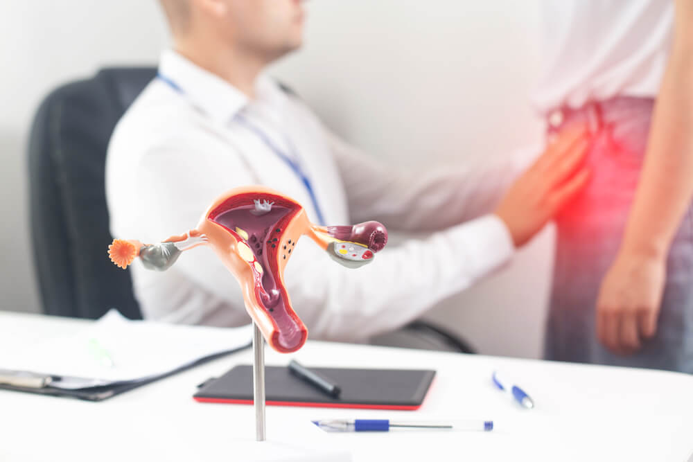
A leiomyoma, or uterine fibroid, is a growth within the uterus that consists of connective tissue and muscle. Also known as myomas, these growths are found in the walls of the uterus. Under normal circumstances, the uterus is usually the size of a lemon. It is in this organ that the baby develops during pregnancy.
Uterine fibroids usually grow as a single nodule. However, in some instances, they can grow as a cluster. Each fibroid cluster ranges in size from about a single millimeter to over 20cm in diameter. In some cases, they can even have a diameter larger than 20cm and be around the size of a watermelon!
Fibroids can either grow within the uterine walls or be found inside the main cavity of the uterus. Sometimes, you might even find them on the outer surface of the organ. Depending on where they are located, fibroids can cause a variety of symptoms that usually differ from woman to woman. Because of how differently they present, it might be difficult to guess where your symptoms might be coming from. Also, because they present differently, the uterine fibroid treatment plan will be different for each individual case.
Causes of Uterine Fibroids
Unfortunately, the exact cause of uterine fibroids is not known. However, they are quite common in women of reproductive age. They are rarely diagnosed in women who haven’t experienced their first period.
Symptoms of Uterine Fibroids
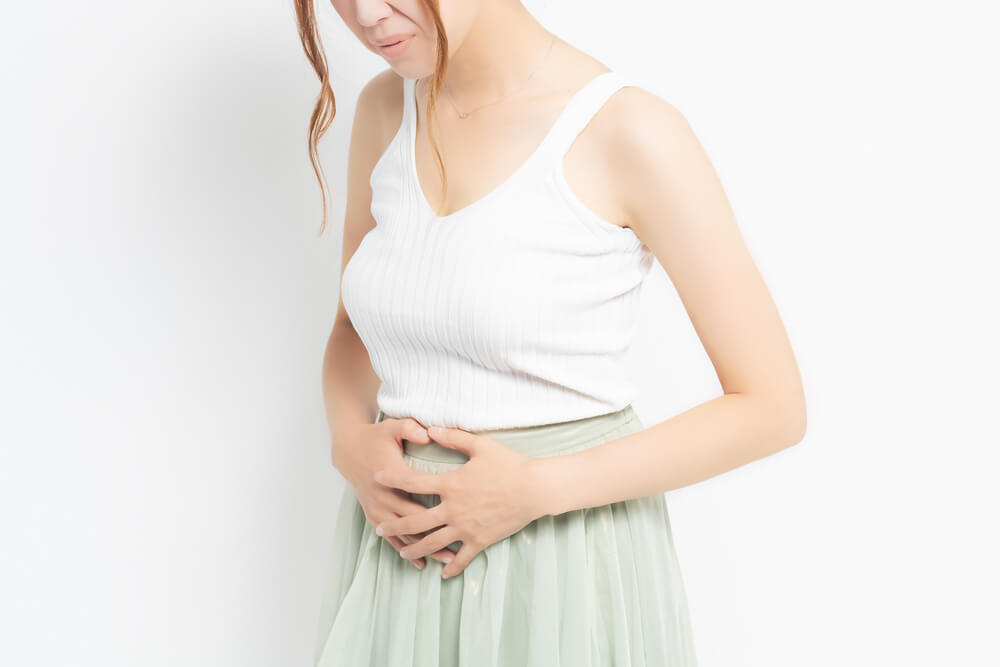
In most instances, uterine fibroids do not display any symptoms. Similarly, they will not require any specialized form of treatment. With asymptomatic fibroids, most doctors will just observe and watch for complications. Asymptomatic fibroids are usually small. However, with larger masses, you might notice the following signs of fibroids:
- Pelvic Pain
- Excessive bleeding during monthly periods
- Bleeding in between periods
- Abdominal bloating
- Frequent urination
- Painful sexual intercourse
- Pain in the lower back region
- Constipation
- Continuous vaginal discharge
- Abdominal distention
It’s been observed that the signs of fibroids, including fibroid pain, usually go away when women reach menopause. This is likely due to the lower levels of circulating hormones, which causes fibroids to shrink.
Location of Uterine Fibroids
Fibroids grow in several different locations in the uterus. Some grow on the inside, while others will be found on the outer surface. It’s important for the doctor to determine the location of fibroids so they can formulate an appropriate treatment plan. This is because the treatment method will depend on the fibroids’ number, size, and location. There are several terms used to describe where the fibroids are located in the uterus and how they are attached.
Submucosal Fibroids
Submucosal fibroids are located inside the uterine cavity within the uterine lining. This is the location where the baby grows when a woman is pregnant. These growths typically attach to the wall of the uterus and extend inwards into the cavity. Submucosal fibroids often cause heavier periods or irregular bleeding.
Intramural Fibroids
These fibroids are located within the wall of the uterus. Instead of extending into the uterine cavity, the intramural fibroids grow inside the muscular wall of the uterus.
Subserosal Fibroids
A subserosal myoma is located on the outer surface of the uterus. These are similar in attachment to submucosal fibroids, with the only difference being that they are on the outside instead of the uterine cavity.
Pedunculated Fibroids
Pedunculated fibroids are not very common. Like subserosal fibroids, they are also located on the outer surface of the uterus. However, unlike the other types, their connection to the uterus is quite loose. They are only joined to the uterine wall by a thin stem or stalk. As a result, they are often seen as mushroom-like growths when using imaging systems. This is because of the thin stalk and a wider top.
Diagnosis of Uterine Fibroids
Because often do not cause any symptoms, fibroids are usually incidentally discovered during routine pelvic or prenatal examinations. However, they may be suspected if you are experiencing pelvic pain or heavy bleeding during your periods. There are several tests that can be performed to diagnose fibroids including pelvic ultrasound, CT scan, or MRI.
Ultrasonography
This is a non-invasive imaging technique used by doctors to obtain pictures of the internal organs using sound waves. The test can either be performed transvaginally or via the transabdominal route. It is often the best and preferred way to image the uterus and ovaries.
Computed Tomography (CT)
Commonly known as a CT scan, a computed tomography makes use of X radiation to come up with detailed pictures of internal organs. This type of scan produces images from several angles. Fibroids are often found incidentally on CT scans. If this is the case, a pelvic ultrasound is often ordered in order to obtain better images of the uterus and ovaries specifically.
Magnetic Resonance Imaging (MRI)
Magnetic resonance imaging uses magnets and radio waves to develop detailed images of internal organs. This is another test used for the diagnosis of uterine fibroids. It is often used if multiple large fibroids are present on a pelvic ultrasound and further imaging of the bladder, colon, or rectum is needed.
Treatment of Uterine Fibroids
Unfortunately, there is no one specific method used to treat fibroids. In fact, in most instances, if the fibroids are not causing any symptoms, the doctor will often recommend watchful waiting. However, if the fibroids are causing problems such as pelvic pain or heavy periods, they can be treated medically or surgically.
Medications
There are several medications that can be used to treat fibroids. One type is Gonadotropin-releasing hormone (GnRH) agonists. This type of medication inhibits the production of the hormones estrogen and progesterone. As a result, menstruation stops, resulting in the shrinkage of the fibroids. While this sounds like an ideal treatment, it is only intended for short-term use (several months) due to side effects. You should talk to your doctor regarding these medications to see if you are a candidate.
Doctors can also use a progestin-releasing intrauterine devices, or IUDs, such as Mirena, Kyleena, or Liletta. These devices can put an end to the excessive bleeding that occurs as a result of fibroids. While these devices do not cause the fibroids to shrink, they often significantly improve the amount of bleeding caused by fibroids.
There are also non-hormonal medications that can be used. These include tranexamic acid, which helps to ease heavy menstrual periods. Doctors can also recommend oral contraceptives, but only to control menstrual bleeding without attacking the actual fibroids. For some, these medications work very well and patients are able to significantly improve their symptoms.
Surgery
There are several surgical techniques that are used to treat fibroids. These include methods such as hysteroscopy, laparoscopy, uterine artery embolization, and hysterectomy.
Hysteroscopy
Hysteroscopy is a minimally invasive technique in which a doctor inserts a thin, flexible tube with a camera on the end into the uterus. This tube is inserted through the vagina, and it’s quite efficient in removing fibroids that are attached to the inner wall of the uterus.
Laparoscopy

Laparoscopy is a minimally invasive procedure commonly used to diagnose abdominal conditions. It is similar to hysteroscopy in that it uses a thin tube with a camera to take a closer look at internal organs. However, with laparoscopy, a thin incision is made in the base of the belly button to pass the camera. With this method, a myomectomy, or removal of fibroids can be performed. Laparoscopy is ideal to remove fibroids that are within the uterine wall (intramural), or stuck on the surface of the uterus (subserosal). Often these surgeries are best performed by a minimally invasive gynecological surgeon or even a gynecological oncologist.
Uterine Artery Embolization
This procedure if performed by a radiologist where small embolic agents to cut off the blood supply are placed through the lower leg veins. These agents reach the fibroids and cause them to shrink. This procedure is only meant to be performed on patients who do not desire pregnancy in the future.
Hysterectomy
Hysterectomy is a surgery in which the entire uterus and cervix are removed (not necessarily the ovaries). A hysterectomy can be performed either through the vagina, by laparoscopy, or with a large incision (like a C-section). This surgery is the most definitive way to treat fibroids as they all are removed at the same time as the uterus. While this is the most definitive way to remove fibroids, it is sometimes a last resort as medications can often work to treat fibroids.
Uterine fibroids are quite common. It’s important to know how they present so you can quickly alert your healthcare provider if you notice any symptoms. However, they can also be asymptomatic, in which case they won’t likely cause any problems. Contact us if you suspect that the symptoms you are experiencing are due to uterine fibroids.