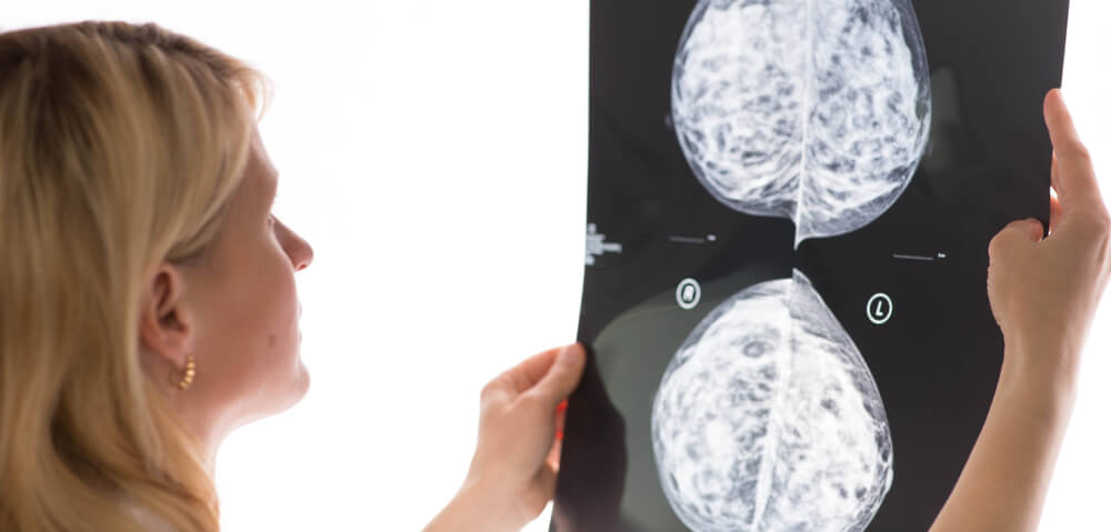
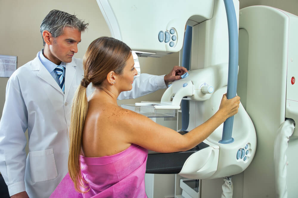
A Breast MRI, short for Magnetic Resonance Imaging, is an advanced medical imaging technique used primarily to detect breast cancer, especially in women who have high-risk or dense breast tissue. This radiology test provides in-depth images of the breast tissue, helping to distinguish between benign (non-cancerous) and malignant (cancerous) areas. It’s often used when other testing procedures like mammogram or breast ultrasound images are inconclusive.
This imaging method utilizes strong magnetic fields and radio waves to create comprehensive images of the breast. One of the key benefits of a Breast MRI with contrast is its excellent soft-tissue image contrast, surpassing the ability of other imaging techniques like CT or X-ray. Thus, it can reveal the presence of small cancers that other imaging methods could miss.
Understanding the importance of Breast MRI is essential in medical health. Specifically, a Breast MRI plays a critical role in breast health as it opens the door for early and accurate detection of potential breast diseases. At Breast Care Center Miami, we prioritize advanced diagnostics for optimal breast care.
Due to its high level of sensitivity, a Breast MRI is quite proficient in detecting tiny lesions or early-stage cancers, enhancing the chances of successful treatment. Its potential to differentiate between scar tissue and recurrent tumors can also significantly aid postoperative follow-ups. This makes MRI for breast cancer an undeniably vital tool in the progressive stride towards robust breast health.
Comprehensive Description of Breast MRI Procedure
The process involved before, during, and after a breast MRI
Before a Breast MRI, patients might receive a contrast dye intravenously to improve image quality. This dye makes specific tissues or abnormalities more visible. Women may need to adjust their mammogram screening appointment to the second week of their menstrual cycle for best results, as hormonal fluctuations can impact the imaging outcome.
During a Breast MRI, the patient lies face down on a padded scanning table, with their breasts placed into a hollow depression in the table, designed for comfort. The table then glides into the MRI machine, where magnetic fields and radio waves develop image layers of the breast tissue. These images are generated without radiation, making MRI Breast Screening a safer alternative.
The intravenous line is removed upon completion of the scan, which typically lasts 30 to 45 minutes. The gathered images are then examined by a radiologist proficient in breast imaging. In cases of detected anomalies, the experts may suggest further evaluation like MRI-guided breast biopsy to confirm whether the irregularity is cancerous.
The types of equipment used and why they are important
The main instrument used in a breast MRI is a magnetic resonance imaging scanner. This creates a strong magnetic field around the patient and sends pulses of radio waves from the scanner. These radio waves produce a three-dimensional representation of the breast, providing more precise details than traditional 2D imaging used in 3D Mammography or breast ultrasound imaging.
Contrast-enhanced Mammography is a recent technique that involves injecting the patient with a contrast agent. This agent helps to highlight blood flow in potential tumor sites during the MRI. This equipment can be especially beneficial when examining dense breast tissue that might previously have been difficult to read.
A coil is another critical component in a Breast MRI. This device is placed around the breast area to boost the quality of the MRI images, enhancing the accuracy of Breast Cancer Detection. In the case of Bilateral Breast MRI, a unique coil that images both breasts simultaneously is typically used.
A high-resolution computer screen to view and analyze the images and special software dedicated to enhancing radiology breast imaging completes the essential equipment. All these tools together contribute to a Diagnostic Breast MRI’s improved efficacy, which is critical for the early detection and treatment of breast cancer.

The Role of Breast MRI in Diagnostic Procedures
Cases where breast MRI is a preferred diagnostic tool and why
While a mammogram screening is a standard procedure for detecting breast abnormalities, a Breast MRI holds a distinct edge in certain situations. When traditional methods such as Mammography or breast ultrasound imaging yield indecisive results, a Breast MRI is often utilized for its ability to generate more comprehensive and distinguishing images.
Given its sensitive nature, a Breast MRI proves to be of immense value while screening women with high-risk breast cancer, primarily due to genetic mutations or family history. It is also chosen as a preferred diagnostic tool to scan dense breast tissue, as it differentiates between harmless and harmful lumps within such an area. Moreover, this imaging technique is quite fitting for identifying the extent of cancer after a new diagnosis and guiding surgical or radiation planning.
Role in early detection of breast disease or condition
Early detection of breast diseases or conditions is crucial for a higher success rate in treatment. Here, the significance of Diagnostic Breast MRI shines through. With its proficient Radiology Breast Imaging, it increases the rate of breast cancer detection, even at the early stages.
Consistent MRI Breast Screening, particularly in cases with a high risk of breast cancer, aids in identifying anomalies when they are minuscule, increasing the chances of successful treatment. The ability of an MRI to characterize and evaluate an anomaly in detail also aids in carrying out a precise MRI-guided breast biopsy, eliminating chances of error and ambiguity.
Moreover, advancements such as Bilateral Breast MRI have led to the simultaneous examination of both breasts, boosting early detection, especially in tumor cases. Especially for women with high breast density, a Breast MRI is valuable as it can reveal cancer that might be hidden during a mammogram screening.
Hence, the role of Breast MRI in the early detection of diseases is proving to be of exceptional importance, supporting healthcare professionals in proactive disease management and improving the patient’s prognosis.
Varied Implications of Breast MRI Results
Interpretation of the outcomes and potential findings
Interpreting the results of a Breast MRI involves a radiologist examining the images generated from the scan for any signs of irregularity. Any abnormality, such as a lump or distortion in the structure of the breast tissue, will be identified and noted. Thanks to the ability of MRI technology to capture images from various angles, it provides a more detailed view, often exceeding that of other imaging modalities such as 3D Mammography or traditional Mammogram Screening.
Common findings during a Breast MRI can range from benign conditions such as cysts and benign tumors to potentially malignant findings that might indicate breast cancer. MRI can also reveal dense breast tissue and conditions like mastitis or inflammation.
Findings are generally categorized into three types:
- benign (non-cancerous)
- probably benign, typically having less than a 2% risk of malignancy
- suspicious
A suspicious result may necessitate further investigation, often an MRI-guided Breast Biopsy, to accurately determine the likely malignancy.
Possible next steps after a breast MRI, based on different results scenarios
If the result of the Breast MRI is benign, no immediate action is required, with the patient usually recommended to continue with routine screenings and mammograms. Probably benign results may warrant short-interval follow-up MRI Breast Screening to monitor the findings. The clinician often guides this decision, considering various factors, including the patient’s health, age, and family history.
When the breast MRI results are suspicious, a biopsy follows to determine whether the detected anomaly is cancerous. Modern techniques like MRI-guided Breast Biopsy are used for precision. Following a positive confirmation of cancer, a multidisciplinary team develops a tailored treatment plan. This might include surgery, radiation therapy, chemotherapy, hormonal therapy, or a combination of these.
In cases such as Bilateral Breast MRI, where both breasts have been evaluated, the following steps are charted out based on findings in either or both breasts. This wide range of potential outcomes underlines the significance of Breast MRI in diagnostic Radiology Breast Imaging. By enabling early Breast Cancer Detection, optimizing treatment plans, and monitoring improvements, Breast MRI significantly contributes to mounting a successful fight against breast ailments, in particular, cancer.
Necessity and Benefits of Breast MRIs
Medical technology advancements have always played a pivotal role in enhancing healthcare services, with Magnetic Resonance Imaging technology among the top game-changers. The application of MRI in the domain of breast health, particularly with Breast MRIs, has made substantial strides in the path of early and precise breast disease detection.
With its superior image quality surpassing mammogram screening, ultrasound imaging, and even 3D Mammography, a Breast MRI is an invaluable tool for clinicians. Whether it’s detecting small lesions among dense breast tissue, monitoring high-risk individuals, guiding biopsies with precision, evaluating the extent of identified cancer, or differentiating between harmless and harmful anomalies, the versatility of this imaging technique is impressive.
Yet, the ultimate value of such a powerful tool rests in its timely and accurate use. This is where the onus shifts to us, the recipients of healthcare services. Taking care of our health should be prioritized, and breast health is no exception.
Adopting routine medical check-ups, specifically after age 40, or as recommended due to specific risk factors, is a small step with a considerable impact. While self-examinations are helpful, they are not replacements for professional screenings and mammograms or the detailed images provided by a Breast MRI.
Remember, the early detection of abnormalities increases the chances of successful treatment. Thus, if a healthcare provider advises a Breast MRI, we must understand the rationale behind this recommendation. Such diagnostic tools are not just about identifying a disease; they also provide us with a more straightforward path toward successful treatment and a healthier future.


