

An abdominal or transabdominal ultrasound helps healthcare providers to visualize internal structures in the abdomen through a low-risk, non-invasive method. This diagnostic technique has many applications and can provide a safe alternative to other imaging modalities.
Continue reading this post to find out how you could benefit from this technology and connect with your local health care providers at Pembroke Pines Ultrasound to receive specialized guidance and care.
What is a Transabdominal Ultrasound?
It is almost as simple as it sounds: “trans-” means “through,” and “abdominal” refers to the abdomen; therefore, any diagnostic that utilizes an ultrasound probe to send signals through the abdominal wall can fall under this category. But what signals are created, and what is this type of ultrasound used for? Let’s dig deeper into how this technology works and why it’s considered an ideal imaging modality for many applications.
How Ultrasound Works

First, you should know that high-frequency ultrasound waves can be produced using a type of transducer called an “ultrasound probe.” (If you have ever seen an ultrasound before, this is the wand that sonographers use to rub on the skin.) Transducers can change one form of energy into another. In this case, the ultrasound probe changes the pressure from the sound waves into electrical signals and vice versa.
So, the sound waves pass through the skin and travel through the body until they meet internal tissues in the abdomen. Then they bounce off these tissues and return to the ultrasound probe, which converts them into electrical signals. This process helps create a picture or moving image, which can then be assessed by a physician or sonographer trained to interpret the data gained from the ultrasound.
Why Ultrasound is Done
Now that you have gained a glimpse into how this technology works, you may be wondering what symptoms you could have that would indicate the usage of this procedure in the first place. Well, the abdominal ultrasound can help diagnose many complications stemming from issues in the abdominal area. These can include the following:
- Abdominal Pain
- Blunt Trauma (injury) to the abdomen
- Hypotension (low blood pressure)
- Hematuria (blood in the urine)
- Fetal ultrasound (for viewing the fetus in pregnancy)
- Vaginal Bleeding
During pregnancy, regular screenings are advised to monitor the developing baby’s health. Examinations for abdominal pain or blunt trauma include the evaluation of fluid, swelling, or masses present in organs such as the gallbladder, digestive tract, liver, pancreas, and kidneys. In the case of hypotension, blood vessels like the inferior vena cava and aorta are screened for infection and improper blood flow. If symptoms include hematuria, health providers scan for kidney stones, swelling, or masses in the genital and/or urinary structures. Finally, for vaginal bleeding, screenings may scan for pregnancy, ectopic pregnancy, or any other uterine issues.
Please contact us if you are experiencing any of these symptoms or complications. At Pembroke Pines Ultrasound, we are experts in this area and have been improving patient outcomes for years.
Ultrasound Versus Other Imaging Modalities
Note that there are many types of imaging modalities, including X-rays, MRI, and CT scans.
A transabdominal ultrasound is often the first-line imaging technique for viewing abdominal structures. It does not expose patients to radiation, is more cost-effective, and is easier for patients and providers to access.
If results are inconclusive or further diagnosis is needed, in some cases, a CT scan or MRI may be ordered as a secondary imaging method. Medical radiation via an X-ray scan would not even be indicated for the aforementioned symptoms. This is because the soft tissues in the abdomen cannot be visualized using X-rays.
Though less common, sometimes an ultrasound with contrast is performed. The contrast solution is not necessary for most procedures. When used, it is delivered through injection and is often less allergenic than other contrast dyes. This is because it primarily relies on microbubbles to fill the blood vessels and organs, providing a more precise image than we would see without the contrast agent.
What to Expect During an Abdominal Ultrasound Procedure
Rest assured that a trained provider or registered diagnostic medical sonographer will be responsible for performing the procedure, guiding you through the process, and interpreting the results. Before the procedure, you should receive further instructions on how to prepare. The provider will discuss your unique case, why the ultrasound is requested, what information will be collected, and how the results can be used to improve your medical care.
The instructions given to you by your provider may differ depending on which structures will be examined. For gallbladder imaging, you may be asked to refrain from eating to improve visualization. On the other hand, if you receive a uterine ultrasound, you may be asked to drink fluids to maintain a full bladder.
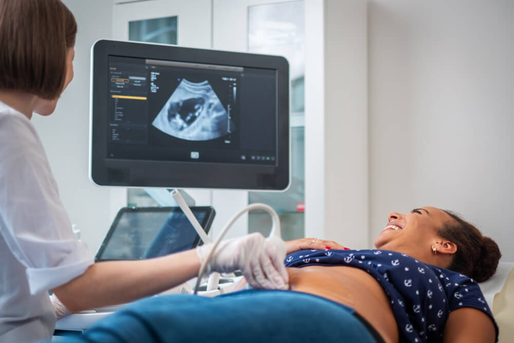
On the day of the appointment, when you enter the patient room, you will lay down with your abdomen facing up. The edges of your clothes and/or dressing gown will be covered with a towel, and ultrasound gel will be used on the probe. Typically, the procedure is performed in a dim environment to allow for better visualization. Throughout the process, you may periodically be asked to take a deep breath and hold it. This can adjust the position of your liver and your gallbladder beneath, which may help with obtaining an image.
Due to the extreme safety and efficacy of the ultrasound, you may immediately resume your regular activities such as driving and exercise, unless otherwise directed by the provider.
How are Ultrasound Scans Safe?
Ultrasound imaging has been utilized in the medical field since the early 1940s, has significantly developed since then, and has been proven to be safe overall. As mentioned before, another benefit of this technology is that it does not expose the patient to ionizing radiation. However, you may note that the sound waves implemented by this technology expose the body to low heat energy and may produce tiny gas bubbles in the process.
Science has yet to prove any long-term effects due to these slight changes. Therefore, healthcare professionals are encouraged to employ “As Low as Reasonably Achievable” practices (ALARA) outlined by the Food and Drug Administration (FDA) and supported by the Center for Disease Control and Prevention (CDC). These guidelines keep patients and healthcare providers safe and are one of the main reasons why diagnostics are not used for recreational purposes but are subject to an indicated medical need.
To determine whether you need an abdominal ultrasound, you must be connected to a health care provider focusing on women’s care at every stage of life. Diagnostic screening remains important for identifying and treating complications that develop early on. Therefore, you must maintain your health through regular appointments and checkups. If you are located in the area and are ready to take charge of your health, contact Pembroke Pines Ultrasound today to find out how we can partner with you on your journey.