
Stereotactic biopsies are a subtype of biopsies that help diagnose cancer cells in the tissue of the breast. Sometimes called a biopsy mammogram, the procedure utilizes a mammography machine using two images of the breast tissue to take a tissue sample for testing.
As you may know, Dr. Eldredge, Dr. Shapiro, and their expert team excel in the diagnosis of breast diseases in Wellington, FL. To help you learn more about the diagnostic processes, the experts at Advanced Surgical Physicians have created this comprehensive article that discusses a specific breast biopsy procedure called stereotactic breast biopsy.
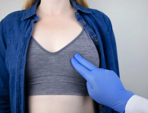
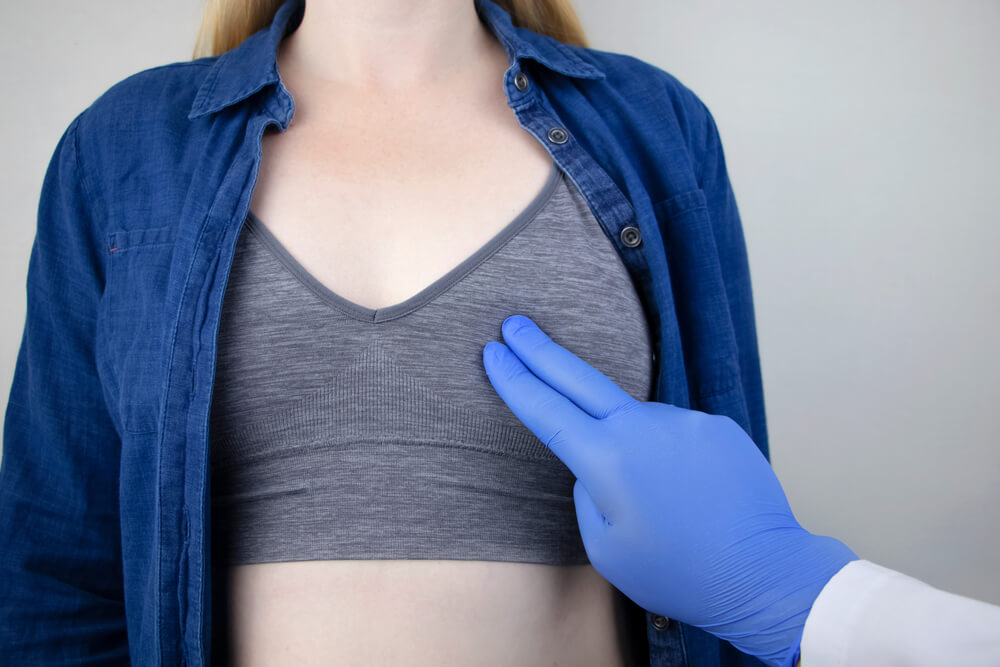
The Importance of a Breast Exam
As you may know, the best and most efficient way to treat cancer is to catch the disease at the earliest possible stage, when the body reacts to treatment most effectively.
On that end, various screening methods can help detect any changes or lumps that may be cancerous. Once these tests return positive, usually, a biopsy is a next step to confirm the diagnosis. And during a biopsy, healthcare providers remove a small tissue sample from the suspicious area for further examination to confirm the presence of mutated cells.
Nowadays, providers use modern imaging technology to help guide the biopsy. Mostly, they use ultrasound, stereotactic breast biopsy, or biopsy mammogram, which uses mammogram technology to guide the needle.
Stereotactic Breast Biopsy

Sometimes referred to as a biopsy mammogram, this technique uses specialized mammography devices that provide guidance when pinpointing suspicious areas within the breast. The machines help by providing X-rays from two distinct angles.
These two sets of images help the radiologist navigate the concerning areas as they remove tissue samples. Then, after the biopsy, a pathologist will conduct further analysis to confirm whether cancer is present.
Usually, stereotactic breast biopsy is recommended when traditional mammograms and other breast examination methods have found:
- A suspicious lump
- Abnormal breast tissue changes
- Small calcium deposits that may signal the presence of cancer
- Suspicious changes to an area where previous surgery was performed
- Breast structure irregularities
A stereotactic breast biopsy may help establish a proper diagnosis when other diagnostic methods have failed to confirm the presence of cancerous cells fully.
What to Expect?
Before undergoing this procedure, you will have a thorough discussion with the provider, who will ask about the medications you use and the supplements you may take (including OTC medications, mineral supplements, vitamins, prescription drugs, or herbal remedies). Also, the doctor will probably ask whether you are pregnant.
Generally speaking, experts will usually recommend the following tips when preparing for a biopsy:
- Wear comfortable clothing before the procedure.
- Avoid using perfume, talcum powder, or deodorant near the breast and the armpits on the day of the biopsy.
- Leave your jewelry at home.
- Remove your glasses, dental appliances, and anything else that may interfere with the X-ray.
Equipment for Stereotactic Breast Biopsy
As said before, this procedure uses X-rays to examine the suspicious area. The design of the mammography equipment ensures that the rays only reach the area that should be examined.
More specifically, the machine can compress, move, and hold the breasts in different positions, allowing experts to take detailed images from different angles.
Stereotactic needle biopsy creates detailed images and helps radiologists get the needle in the right area before taking tissue samples.
On that end, stereotactic needle biopsy uses different needle types to collect breast tissue samples:
- VAD or vacuum-assisted devices to suck out several samples with a single insertion
- CN or core needle, which collects a single sample per insertion
During the Procedure
In most cases, these biopsies are performed as outpatient interventions, with patients being awake throughout the biopsy, which takes no more than 30 to 60 minutes.
Most patients experience no pain or just a slight discomfort during the procedure without any scarring to the breasts afterward.
During a stereotactic needle biopsy, the patient will lie face down on a prone table with an opening for the breasts or sit upright, facing the mammography machine.
Then, the radiologist will numb the area using a local anesthetic. After that, they will make a tiny cut on the breast skin for the biopsy needle. The biopsy needle is then steered through the incision and directed to the targeted areas. At the same time, the process is coordinated with the help of two images generated by the mammography machine. When the needle is in place, the radiologist will obtain more images to ensure the needle is in the right place and collects the necessary tissue.
After this is all done, the provider will remove the needle and place a marker on the area where the procedure was performed for future reference. Lastly, they will take some more images with the mammogram.
After the Biopsy
When all is done, the provider will dress the area with sterile strips and may apply pressure to the site to prevent or stop bleeding.
Following the procedure, patients may experience some soreness, bruising, and swelling, which can be quickly resolved with simple ice packs and OTC pain relievers.
Still, if patients experience draining of fluid, severe bleeding, swelling, heat, and redness in the area, they should reach out to their doctor as soon as possible.
Lastly, following the appointment, patients should avoid vigorous exercise and other strenuous activities for at least a day.
Discussing the Results

Typically, a pathologist will be responsible for examining the collected tissue samples. They will also determine whether cancer cells are present in the gathered samples.
After establishing a diagnosis, they will let the doctor who ordered it know the results. Then, this healthcare provider will be responsible for sharing the results with the patient and discussing treatment options if necessary.
Doctors can gather plenty of information from these tissue samples. Apart from establishing the presence of cancer cells, biopsies may also show what kind of cell mutations triggered the disease in the first place and whether the cells are hormone receptor-positive. These things can help doctors and patients develop individualized treatment plans to ensure the best possible outcome.
Risks
Unfortunately, stereotactic breast biopsies involve some minor risks, which include the following:
- The patients may experience some pain.
- In rare cases (less than one percent), hematomas may develop in the area where the biopsy was performed.
- In rare cases, the area may get infected.
- In even rarer cases, the needle may go through the chest wall, causing complications.
- The procedure can’t be performed on pregnant women as the radiation may harm the baby.
- The procedure may be challenging to perform if the patient has small breasts.
Take Advantage of Modern Technology
In the past, more invasive surgery was the only option for breast tissue biopsy. Fortunately, with modern technological advancements, experts can now perform biopsies with minimally invasive techniques more quickly and with faster results.
That said, if you have any concerns or questions about this type of biopsy or breast cancer in general, feel free to contact our expert team today.
Please remember that the purpose of this article is strictly to provide information. You should contact your healthcare provider or our office right away if your symptoms persist or return.


