
Pregnancy is a remarkable journey filled with anticipation and wonder. As expectant parents, we eagerly await the arrival of our little one, often finding solace in the rhythmic thumping of their developing heart. But what if there was a way to gain a deeper understanding of that tiny heart’s rhythm and structure? This is where fetal echocardiography comes into play, offering an incredible window into your baby’s cardiac health. After all, doing an echo scan during pregnancy at a reputable pregnancy care center in Plantation, Florida, is likely the optimal approach to initiating healthy prenatal support. In this article, we’ll dive into the world of fetal echocardiography, exploring its purpose, procedure, significance, and more.
What Is Fetal Echocardiography?
Fetal echocardiography, often referred to as a fetal echo, is a specialized ultrasound technique that focuses on imaging the developing heart of a fetus. Just like traditional ultrasound, it uses sound waves to create images, but the difference lies in the level of detail and focus. Fetal echo tests provide an intricate view of the baby’s heart structures, allowing healthcare professionals to detect potential heart defects and irregularities long before birth.
When Is Fetal Echocardiography Necessary?
Fetal echocardiography is typically recommended when there are factors that may increase the risk of congenital heart defects or when an anomaly is suspected during routine ultrasound exams. High-risk pregnancies, maternal diabetes, genetic conditions, or a family history of heart issues might prompt a healthcare provider to suggest a fetal echo scan during pregnancy. By identifying potential heart problems early on, medical teams can develop a comprehensive plan to address the baby’s needs even before they are born.
What Are the Risks of Fetal Echo?
The good news is that fetal echocardiography is considered a safe procedure, much like traditional ultrasounds. The ultrasound waves used in the test are non-ionizing, which means they don’t pose any risk of radiation exposure to you or your baby. However, as with any medical procedure, there is a very small possibility of discomfort or allergic reactions related to the gel used during the test. Your healthcare provider will be sure to address any concerns you might have before the procedure.
How to Prepare for the Procedure?
Preparing for a fetal echo is straightforward and generally does not require any special steps. You may be advised to wear comfortable clothing and avoid applying lotions or oils to your belly before the exam. It’s also a good idea to have a list of questions ready for your healthcare provider, ensuring that you have a clear understanding of the process and its potential outcomes.
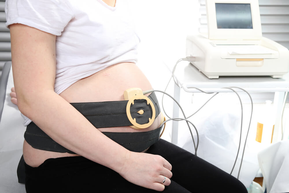
What Happens During the Exam?
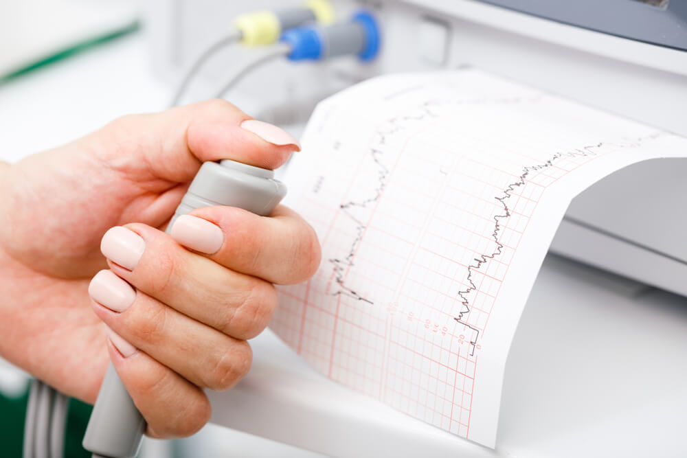
During fetal echocardiography, you’ll likely be asked to lie down while a trained sonographer uses a handheld device called a transducer. This device emits sound waves that bounce off the baby’s heart structures, creating detailed images on a monitor. The sonographer will move the transducer gently over your belly to capture various angles of the heart. The procedure is generally painless and can take around 30 to 60 minutes, depending on the baby’s position and cooperation.
What Happens After Fetal Echo?
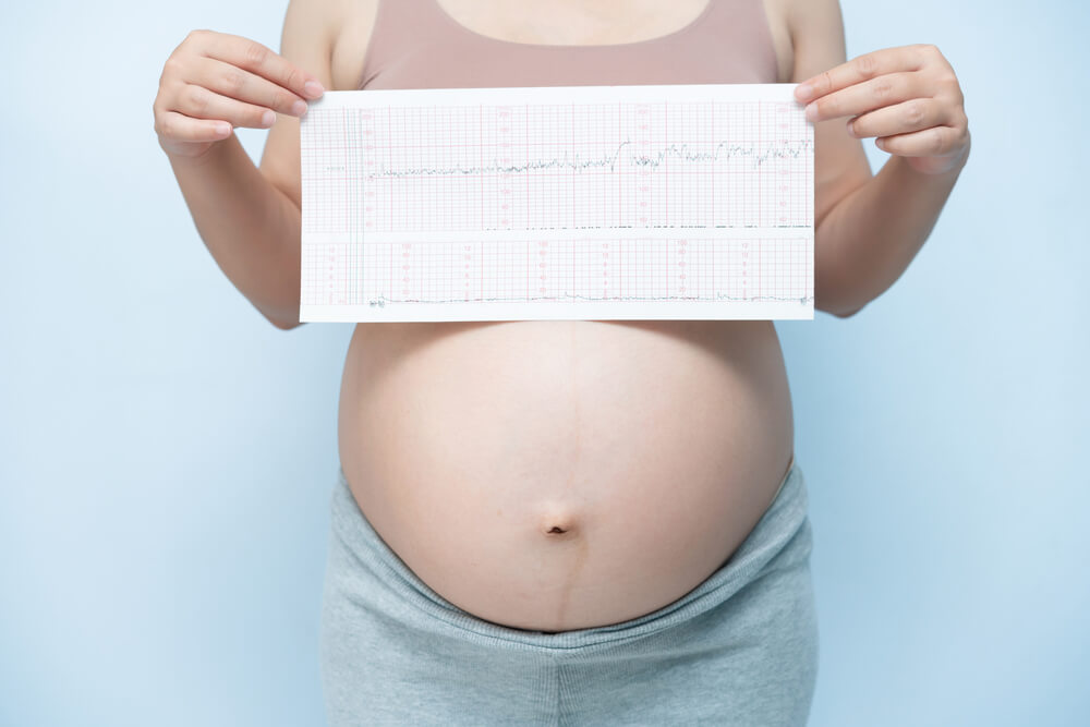
Once the fetal echo test is complete, a specialized team of healthcare professionals, including pediatric cardiologists, will review the images and analyze the baby’s heart structures. If any abnormalities are detected, they will discuss the findings with you in detail, providing a comprehensive understanding of the situation. It’s important to remember that not all anomalies indicate a severe problem; some might resolve on their own as the baby continues to develop.
What Do the Results Mean?
The results of fetal echocardiography can vary widely. In some cases, the baby’s heart may show no abnormalities, offering you peace of mind and reassurance. In other instances, minor irregularities might be detected that could require additional monitoring throughout the pregnancy or after birth. In more complex scenarios, significant heart defects might be identified, necessitating further medical interventions or surgeries after delivery.
What Are the Next Steps?
If a fetal echo reveals any concerns or abnormalities, your medical team will guide you through the next steps. This might include more frequent monitoring, consultations with pediatric specialists, or the development of a comprehensive care plan for your baby’s arrival. It’s important to stay connected with your healthcare provider and ask any questions you may have as you navigate this journey.
Why Is This Test Important?
Fetal echocardiography isn’t just about diagnosing potential heart issues; it’s about giving your baby the best possible start in life. By identifying and addressing heart defects before birth, medical teams can be better prepared to provide timely interventions and care immediately after delivery. This test empowers parents with knowledge, ensuring they are well informed about their baby’s health and any potential challenges that might lie ahead.
Fetal echocardiography not only aids in the early detection of heart issues but also plays a vital role in advancing medical research. The insights gathered from these detailed imaging procedures contribute to a better understanding of congenital heart defects and their underlying causes. Researchers can use this information to develop new treatment approaches, refine surgical techniques, and enhance prenatal care strategies. This continuous cycle of research and refinement ultimately improves outcomes for babies born with heart conditions.
Moreover, the data collected through fetal echocardiography can be anonymized and used for population-level studies. This enables healthcare professionals and researchers to identify trends and patterns in heart defects, leading to improved preventive measures and strategies for reducing the overall prevalence of congenital heart conditions. This collaborative effort between medical practitioners and researchers showcases the multidimensional impact of fetal echocardiography on both individual patient care and the advancement of medical knowledge.
Empowering Parents: Informed Decision Making
Pregnancy is a time of anticipation, excitement, and, understandably, a certain degree of anxiety. Fetal echocardiography not only provides valuable insights into the health of the developing heart but also empowers parents to actively participate in their baby’s medical journey. Armed with knowledge, parents can make informed decisions about their baby’s care, considering various treatment options and potential outcomes. This involvement fosters a sense of agency and partnership with the medical team, ensuring that the best possible decisions are made for the well-being of the baby.
To Conclude
Fetal echocardiography is a remarkable tool that brings the miracle of modern medicine to the realm of prenatal care. Through detailed imaging of your baby’s heart, this procedure can detect and address potential heart issues, allowing for timely interventions and comprehensive care plans. As an expectant parent, embracing this opportunity for early detection can make a significant difference in your baby’s journey toward a healthy life. Remember, your healthcare provider is there for you every step of the way, answering your questions and guiding you through the process with compassion and expertise. That said, you can always count on Broward Complete OB-GYN Wellness Center for complete care and support.