Ultrasound, also known as medical sonography, is a method used in diagnostics to create images from within your body with the help of sound waves. Medical professionals usually use ultrasound to examine soft tissues, follow the development of the fetus, diagnose a condition, or as guidance through some procedures.
The benefits of using ultrasound are numerous – they are painless and non-invasive, the patients are not exposed to any radiation, and they are accessible in most facilities. If you are looking for an ultrasound scan in Doral, FL, make sure you have done enough research to pick the best option.
In the field of gynecology, different kinds of ultrasounds are used to diagnose conditions. People usually consider ultrasounds only when they are utilized to follow the progress of a pregnancy, however, this method is also used to diagnose many different health problems concerning women’s reproductive health.
Different Gynecological Sonography Exams
An ultrasound can examine different organs within the woman’s reproductive system, such as the ovaries, vagina, uterus, cervix, pelvis, bladder, and fallopian tubes. Although the most common, obstetric ultrasounds are not the only exam where gynecologists use an ultrasound to examine women’s health conditions.
The doctors can use an ultrasound to examine and diagnose the following conditions:
- Endometriosis
- Uterine fibroids
- Ovarian cysts
- Pelvic inflammatory disease
- Malformations in the womb
- Issues with fallopian tubes such as blockages
- Tumors
- Infertility issues
- Displacement of an IUD
After this relatively quick and completely painless examination, the doctors can determine the direction of your further treatment and care. Another positive side of ultrasounds is that the medical professional conducting the exam has the information about a condition right away, without a need to wait for the results or get a separate analysis of the exam results.
There are several types of ultrasound apart from the obstetric one, such as the pelvic ultrasound (transvaginal/endovaginal and transabdominal ultrasound), 3D ultrasound, saline ultrasound, and alike.
Pelvic Ultrasound
The pelvic ultrasound is most frequently used when diagnosing gynecological issues. There are three types of pelvic ultrasound where the two types mentioned above are used on women, while the transrectal ultrasound is used only on men.
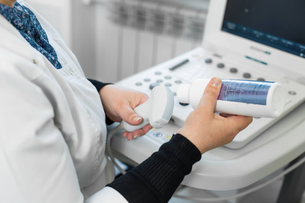
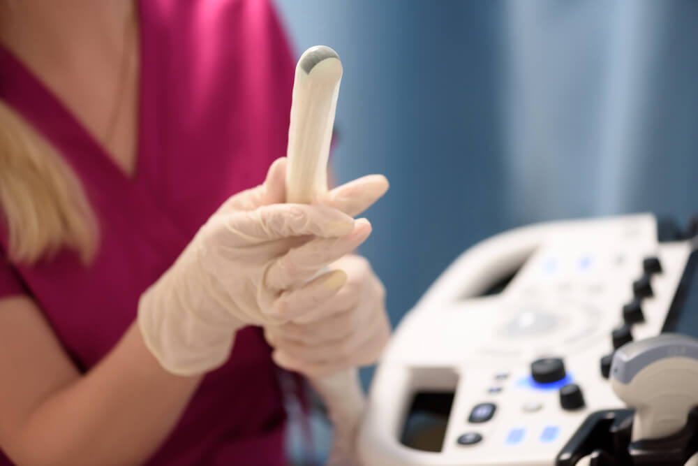
The transvaginal ultrasound examines the female pelvic organs (bladder, uterus, cervix, vagina, and rectum), lower abdomen, and different tissues. During the exam, the doctor inserts a long thin ultrasound transducer into the vagina to take a look at the abovementioned organs.
A specific type of gel or a latex or plastic cover is used for the transducer so it does not cause painful sensations for the patient, but also to prevent any air traps, to help with the movement of the instrument, and to create higher quality images. The patient lays on their back, with their feet in the stirrups.
This type of ultrasound is needed when:
- Locating an IUD
- There is irregular bleeding or issues with your period
- Evaluating a mass in the organs, such as fibroids, cysts, or tumors
- There is pelvic pain
- Diagnosing causes of infertility
- Looking for an ectopic pregnancy
Unlike the vaginal one, the transabdominal ultrasound is done over the surface of the abdominal skin. The patients lay on their back, and a gel is applied over the abdomen to help remove access air between the skin and the transducer and improve the sound waves’ overall quality during the exam.
The 3D ultrasound is a volumetric technology that creates a three-dimensional view of internal structures. Sometimes, patients want to have a 3D ultrasound, mostly in cases when the parents want a more precise in utero image of their infant. Although not crucial, 3D ultrasound is helpful when detecting congenital disabilities and health issues that sometimes cannot be seen through regular ultrasound imaging.
How to Prepare and What to Expect at an Ultrasound?
Generally, there are no special preparation steps before having an ultrasound. The only preparatory measure when you have a transabdominal ultrasound is that your bladder needs to be full, so make sure you drink plenty of water before the examination. On the other hand, if you have a transvaginal ultrasound, your bladder needs to be empty, so do not drink any liquids and go to the bathroom before the exam.
Some doctors might ask you to refrain from eating before the ultrasound, so listen to the directions your chosen medical professional gives you, and you will be well prepared. Also, wear loose clothes to feel more comfortable and remove the clothes easily if needed.
The ultrasound examination does not take longer than an hour, and some may last only up to thirty minutes. During the exam, you are awake, and you will be able to speak with the medical professional conducting the examination. There is no pain during any type of ultrasound examination, but you can feel slight discomfort during the transvaginal ultrasound.
Benefits of the Ultrasound Method
To recapitulate, there are many benefits of having an ultrasound, and below, you can read more about some of them:
- It is non-invasive
- It is not painful in any way, just a little uncomfortable at times
- There is no need for incisions or injections
- It is commonly available, and it is mostly inexpensive in comparison to other methods
- It is very safe because there is no radiation
- It is an excellent choice when examining soft tissue
- The images are created at the time of the examination so that the doctor can have a prompt reaction to any potential conditions and issues
All these benefits make the ultrasound examination method a popular choice in modern medicine. Some conditions also need additional examinations, but the ultrasound is usually the first step in getting a correct diagnosis and treatment.
So Are There Any Risks?

There are no known risks to ultrasound examinations. Numerous studies have shown that there are no risks involved with having an ultrasound, be it the effect of the sound waves or the exam itself.
The downside of the ultrasound exam is that sometimes this particular imaging technique is disrupted due to air in the organs, density of the bones, and alike. This said it can still be successfully used to detect fluid around organs, such as lungs, or fractures and infections around bones or other organs.
Schedule an Appointment
An ultrasound examination is one of the most accessible imaging methods, but it might still be a stressful experience for you. We are here to make it as comfortable as possible, so if you are planning on having a gynecological sonogram, feel free to contact us, and we promise we will make your experience as enjoyable as possible.
Schedule an appointment online, or just call us! Our team is here to walk you through the whole process without stress. We are here for you.