Diagnostic radiology is a branch of medicine that uses non-invasive scanning to determine what is happening inside your body and diagnose illnesses and injuries. Diagnostic radiology can create images of your organs, bones, soft tissues, blood vessels, muscles, and many other internal structures that aren’t visible to the naked eye.
Diagnostic radiologists specialize in diagnosing and treating health conditions using a range of imaging equipment, including X-rays, ultrasounds, CT scans, and MRIs. Diagnostic radiologists often specialize in specific areas of radiology as well, such as breast imaging, oncology radiology, cardiovascular radiology, or pediatric radiology.
TopLine MD Alliance Network diagnostic radiology centers are equipped with the most modern technology, enabling them to deliver high-quality, high-precision results and provide a comfortable experience for all patients. Learn more about diagnostic radiology, how it works, what to expect at a diagnostic imaging appointment, and more.
What types of tests and scans are included in diagnostic radiology?
Diagnostic radiology includes many types of tests and scans that have one primary purpose: to view the inside of the body. Yet they often use different technologies to create the images, such as radiation, sound waves, radio waves, and magnetic fields.
Some common types of diagnostic radiology tests and scans include:
X-ray
X-rays, the oldest form of diagnostic radiology, send a small amount of radiation through the body. The radiation passes through soft tissues but is absorbed by dense materials like bones and joints – creating an image where bones and joints appear white and soft tissues appear shadowy.
X-rays are used to detect conditions like:
• Bone fractures
• Foreign objects in the body
• Tumors or masses
• Pneumonia in the lungs
• Other infections
Ultrasound
Ultrasound uses high-frequency sound waves to capture images. A wand-like device called a transducer sends the sound waves into the body, which reflect and are converted into real-time images by a computer.
Various tissues reflect the sound waves differently, creating shades of gray and white on the ultrasound images. The images can be static, in motion, or converted into 3D or 4D views with the proper equipment.
Ultrasound has many key uses, including:
• Pregnancy (confirming pregnancy, monitoring fetal heart rate and growth, checking for abnormalities, and more)
• Finding the source of pain, swelling, or infection
• Diagnosing injuries like strains or sprains
• Monitoring blood flow through the heart and blood vessels
• Evaluating how organs function
CT scan
Computed tomography (CT) scans use X-rays from multiple angles to create cross-sectional or 3D images of the body, which provide more detailed views of organs, bones, blood vessels, and other tissues.
The patient lies inside the CT scanner as it rotates around them, taking images. For certain CT scans, a contrast dye may be swallowed or injected into a vein to help certain organs or blood vessels show up more clearly.
The actual scanning time is usually short, but the entire process may take 10-30 minutes or more. It’s important to remain as still as possible during the CT scan, as motion can blur the images.
CT scans are used to detect conditions like:
• Complex bone fractures
• Blood clots
• Internal bleeding
• Tumors or masses
• Kidney stones
• Appendicitis
• Injuries from trauma
CT scans can also reveal how a tumor is responding to treatment, help physicians plan surgeries and radiation therapy, help guide needles during biopsies, and more.
Mammogram
Mammography also uses multiple X-rays from different angles to create a 2D or 3D image of the breast. Standard 2D digital mammography uses two plates to compress breast tissue and takes two images of each breast (one from above and one from each side), while 3D mammography takes a series of low-dose X-ray images as it moves in a small arc around the breast.
For women without any breast cancer symptoms or other issues, a 2D screening mammogram is usually the first step. Women who have breast cancer symptoms or a concerning image on their screening mammogram will likely need a 3D diagnostic mammogram. Diagnostic mammograms are often used to screen women who have been treated for breast cancer in the past as well.
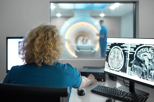
MRI
Magnetic resonance imaging (MRI) uses a powerful magnetic field and radio waves to create detailed images of soft tissues, organs, blood vessels, and more. MRI scans are particularly useful for examining soft tissues that are difficult to see with other forms of diagnostic imaging, such as ligaments, muscles, tendons, and cartilage.
The patient lies on a table that slides into a large, tube-shaped machine. The strong magnets within the machine align with hydrogen atoms within the patient’s body. Radio waves are then sent to the area being scanned, causing the hydrogen atoms to emit signals. A computer receives these signals and uses them to create cross-sectional or 3D images.
An MRI can take anywhere from 20 minutes to two hours, depending on the area being scanned, the number of images needed, and whether a contrast dye is required.
MRIs are used to detect conditions like:
• Strokes
• Brain injuries
• Aneurysms
• Tumors
• Ligament tears
• Pinched nerves
• Spinal cord injuries
• Joint diseases
• Congenital heart defects
• Blood vessel abnormalities
Is diagnostic imaging safe?
Yes, diagnostic imaging is considered safe, as the benefits of accurate diagnosis and treatment far outweigh the potential risks.
Today’s imaging equipment is designed to use the smallest possible amount of radiation while still producing a clear diagnostic image. Physicians only recommend imaging when it is necessary to diagnose, monitor, or treat a health condition. Ask your physician about the possibility of ultrasound or MRI testing if you are concerned about radiation use.
Do I need a referral for diagnostic radiology?
Yes, a referral is usually required for insurance companies to cover the cost of diagnostic imaging services. Contact your insurance company to confirm if you need a primary care physician’s referral.
How should I prepare for a diagnostic imaging appointment?
It’s important to follow any specific instructions you receive, such as not eating or drinking or having a full bladder. You must also inform your diagnostic radiologist about any medical conditions you have or if you are pregnant.
Here are some general tips to follow:
• Wear loose-fitting, comfortable clothes – Avoid clothes with metal zippers, snaps, or other hardware. You may be asked to change into a hospital gown. For mammograms, you will have to remove your bra, so consider wearing a two-piece outfit.
• Leave other metal at home – Jewelry, piercings, watches, and other metal items should also stay home.
• Arrive early – Plan to arrive 15-30 minutes before your imaging appointment to check in and fill out any necessary paperwork.
• Be prepared – Bring your insurance card, medication list, and referral form(s) with you.
• Relax – You may be afraid of confined spaces, nervous about the results of your images, or something else. Try relaxation techniques like deep breathing or meditation, and/or bring a support person with you. For MRIs, you can also discuss prescription sedation with your physician in advance.
How and when will I get my results?
It can take anywhere from a few hours to a few weeks to get your diagnostic imaging results, depending on the type of test or scan you are having.
Once the technician takes the images, a diagnostic radiologist will review them and create a detailed report for your referring physician. This process may take longer if the diagnostic radiologist needs to consult with any specialists.
These are the standard timelines you can expect:
• X-rays – X-rays are often the fastest type of imaging, with preliminary results sometimes ready in as little as an hour or two. (The official report from the diagnostic radiologist may take additional time.)
• Ultrasound – Pregnancy ultrasound images are usually evaluated in real time, while other ultrasound results may take up to a week, depending on their complexity.
• CT scans and MRIs – CT scan results are often available within 24-48 hours, while MRI results typically take 1-2 weeks due to the intricacy of the images. Emergency cases receive results much faster, sometimes within an hour or two for CT scans.
For the most accurate timeline, ask the technician or your physician when they expect to have your results. Your physician’s office will likely contact you to schedule a follow-up appointment or discuss your results once they have the diagnostic radiologist’s report.
Find a TopLine MD Alliance Network provider today
The TopLine MD Alliance was created by physicians who came together to make healthcare experiences better for patients. We help patients navigate the healthcare system, connecting them with top-of-the-line healthcare providers, practices, diagnostic centers, and imaging centers we trust.
The TopLine MD Alliance brings together high-quality care and exceptional service while ensuring that you are always satisfied with your choice of medical providers. To get started, find a provider near you today.