With the ever-evolving advances of modern technology, doctors now have more options than ever when it comes to diagnosing a patient’s condition. There are both invasive and non-invasive methods. Other options offer exploratory or minimally invasive diagnosing techniques. Diagnostic radiology is a group of diagnostic methods that use non-invasive techniques to locate and identify certain conditions and diseases. The types of tests and equipment used in diagnostic radiology generally use a low dose of radiation to generate a detailed image of a specific area.
Radiology is essential for diagnosing many diseases and conditions, particularly cancer. It is a more important section of medicine than many people realize. In reality, patients cannot be effectively managed and treated by doctors without diagnostic imaging. Doctors rely on the test results of diagnostic radiology to determine a proper treatment plan for a patient.
What Is Considered Diagnostic Testing?
Diagnostic radiology is a specialization of medicine that involves generating images of the inside of the body through a range of procedures. These procedures can be anything from MRI and CT scans to X-rays and ultrasounds. Diagnostic tests can consist of physical examinations and biopsies to pregnancy tests and cancer screenings. Diagnostic testing is defined as any approach used to collect information in order to make an informed clinical decision about a patient. While the types of diagnostic tests can vary, they all are used for the goal of estimating the probability of a certain disease or condition in a person.
Some of these diagnostic methods require a compound to be consumed or a chemical to be injected to increase vein visibility. Other types of these tests call for the doctor to put you under anesthesia and use a scope to determine the problem.
What Procedures Are Done in Radiology?
There are a number of different diagnostic radiology methods available in the current age of medicine. Types of diagnostic radiology methods include the following:
- Ultrasound
- Computed Tomography (CT) Scans
- Nuclear Medicine Scans
- Radiography (X-Rays)
- Magnetic Resonance Imaging (MRI) Scans
- Coronary Calcium Scoring
- Virtual Colonoscopy
- Fluoroscopy
- Arthrogram
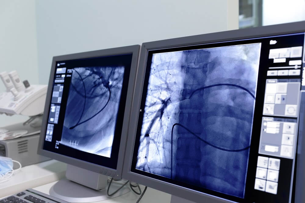
The X-Ray machine is a commonly utilized method of diagnostic radiology. This makes use of x-rays – a form of electromagnetic radiation – to generate images of the inside of the body. X-rays are a non-invasive procedure that offers doctors a helpful look at a condition the patient may be experiencing.
CT scanner machines are another method that utilizes the same equipment as x-rays. These machines provide a cross-sectional look at the body. CT scans are often used when a doctor is looking to create a uniquely detailed image that will allow them to locate the source of the problem. These are especially useful when looking at soft tissue problems and damage.
What Is a Radiology Test?
A radiologist studies for over a decade to learn the ins and outs of examination techniques, anatomy, equipment protocol, and radiation protection to be able to offer the best patient care while on the job.
When a doctor needs to look further than on the outside of a body to see what’s going on, they resort to diagnostic imaging. Diagnostic imaging offers a non-invasive way for doctors to look inside the body. They are able to do everything from track how an organ is functioning and ascertaining the extent of an injury to monitoring joint movement and diagnosing diseases.
What Can Radiography Diagnose?
Diagnostic radiology can be used to diagnose a variety of problems. Diagnostic imaging is able to identify problems ranging from broken bones and blood clots to heart conditions and gastrointestinal conditions.
Diagnostic radiology isn’t just used for identifying problems, however. Doctors can also use diagnostic radiology to monitor the way in which a person’s body is responding to a certain treatment. Additionally, diagnostic radiology can also be used to screen for diseases like colon cancer and breast cancer.
What Can a Radiologist Diagnose?
A radiologist is a doctor that specializes in diagnosing diseases. Radiologists accomplish this using a combination of medical imaging, such as radiology, with procedures like X-rays, CT scans, MRI scans, nuclear medicine, and ultrasounds. They use these procedures and modes of imaging to assess a patient, see inside the body, and diagnose conditions.
A radiologist works alongside your referring physician to prescribe the best plan of action by selecting the proper tests to be performed. The radiologist and their fellow technologists interpret your imaging results, and your radiologist will recommend follow-up tests and possible treatment options.
General practitioners and specialists consult radiologists for the most effective, safest methods of treatment for a patient. In most scenarios, a patient will never come into contact with their radiologist during the diagnosis and treatment process. Instead, the radiologist is working alongside the doctor to help make the best decisions about a patient’s health.
Radiologists work in every field of medicine and specialize in every part of the body. You might run into a radiologist next time your doctors are diagnosing a problem with your heart, lungs, circulatory system, reproductive system, nervous system, urinary system, or breasts. A radiologist is a very important person to have on your side in times of medical diagnosis and treatment.
Interventional radiology is one way of treating and diagnosing certain conditions without the need for scopes or surgery. An interventional radiologist is a highly trained professional who has extra training and experience in using imaging technology such as ultrasounds, MRIs and CT scans to guide medical procedures. Interventional radiology is used in treating liver and kidney problems, blockages in arteries or veins, cancers, and back pain. The patient may be awake or mildly sedated during such procedures.
Is an Ultrasound Considered Radiology?
One of the other most common types of diagnostic radiology testing methods is the ultrasound. An ultrasound is a type of body scan that uses high-frequency sound waves to generate an image. The word “ultrasound” means a sound with a frequency that humans are incapable of hearing. During an ultrasound, these high-frequency noises pass through fluids and soft tissue in the body and bounce off of any denser surfaces it meets. As a result, an image of the inside of the body is created.
This type of testing method is often used to produce images of unborn babies as they are developing. Oftentimes, ultrasounds are also used to produce images of organs, muscles, tendons, blood vessels, and the heart. The resulting image can offer valuable insights into the body and can lead to the diagnosis of many diseases and conditions. Ultrasounds are especially helpful when it comes to problems with the soft tissue within a body.
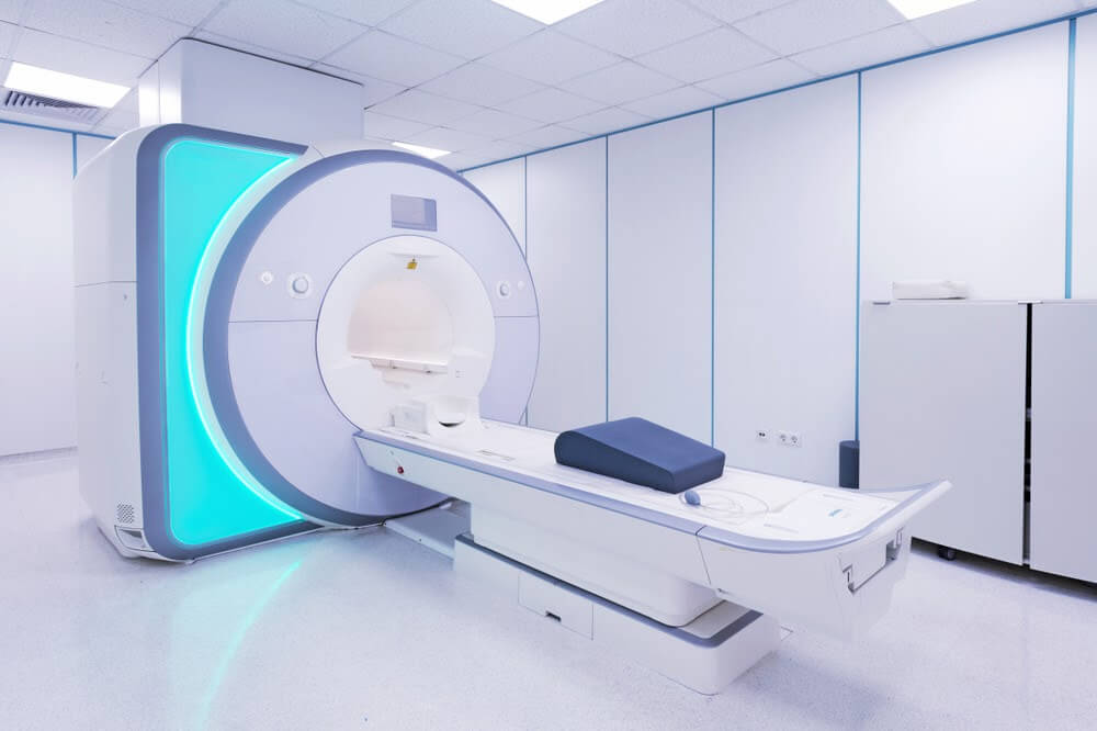
Is an MRI Considered Radiology?
MRI scans are one of the most common forms of diagnostic radiology used for testing. Instead of using radiation, MRI machines work by using a magnetic field to create images of objects inside a body. MRI scans are commonly used to examine a person’s bones and other parts of the body that a CT scanner might have difficulty generating a clear image of. Each image is called a slice, and one exam can produce dozens or even hundreds of images.
There are a number of tools doctors can use to help diagnose and treat patients. From X-rays and MRIs to ultrasounds and CT scans, diagnostic radiology offers physicians a non-invasive way to look at what’s going on in the body. You can visit a TopLine MD affiliated diagnostic imaging center in South Florida, including the Diagnostic Center for Women, Care Diagnostics, Midtown Women’s Center, and Pembroke Pink Imaging. Request an appointment today or ask your primary care provider for a referral.